Taste buds
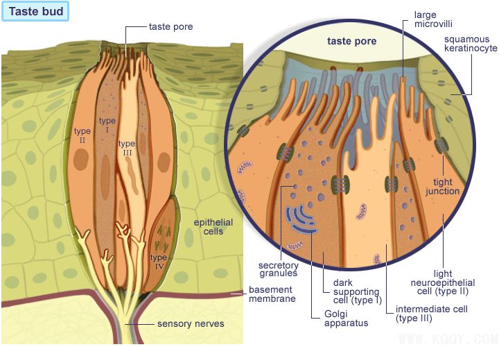
Taste buds: These are pale stained ovoid bodies that extend from the basal lamina, through the epithelium to the surface. At the surface, the squamous epithelial cells form the opening called the taste pore. The microvilli of the columnar cells of the taste bud open into this pore. There are 4 types of cells: three are columnar and extend from the basal lamina to the taste pore, where they end by forming a number of rather large microvilli. They are Type I dark supporting cells, Type II light neuroepithelial cells, and Type III intermediate cells. The nuclei of the columnar cells are placed at their central regions. A fourth cell type, the basal cell, Type IV, is peripheral to the other cells and does not reach the taste pore. It is a stem cell that renews the other cells with a turnover in the order of 10 days. The support cells are secretory and contain secretion granules with a content that resembles the amorphous material surrounding the microvilli in the taste pore. The neuroepithelial cells, 4-20 per taste bud, are dispersed throughout, between the supporting cells. These cells contain vesicles that resemble synaptic vesicles and they are related to the free nerve endings that enter the taste bud through the basal lamina. The other columnar cells may also be innervated. The substances dissolved in the saliva stimulate the sensory taste hairs (the microvilli) and the impulse produced is conducted to the nerve endings. To remove the stimulatory substances, the secretions of Von Ebner glands flow from the bottom of the crypt.
- Long Island College Hospital - [education]
- Faculty of Dental Medicine - H [education]
- The American Association of Or [organize]
- Summer Institute in Clinical D [organize]
- Academy of Osseointegration [organize]
- University of North Carolina a [education]
- American Orthodontic Society [article]
- American Equilibration Society [article]
- Niigata University - Japan [education]
- University of Buffalo [education]