ENDODONTICS AND ESTHETIC DENTISTRY(7)
Pretreatment Radiographs
Reconstructive planning requires a full set of well-angulated long cone-exposed films or digital images using film holders like XCP (DENTSPLY/Rinn, Elgin, IL), which enables 90-degree angulation of the x-ray beam on the film or sensor. In addition to a good angulation on the radiograph, these film holders will enable the operator to take comparable films or digital images over time, which is very important when evaluating healing or failure.
When dealing with extensive cases, panoramic film is equally essential. If the patient requires endodontic therapy and is referred for treatment, these films and a description of the goals and objectives of the referring dentist should be sent to the endodontist prior to the patient's first appointment. Most endodontists will take additional films of the teeth to be treated to establish a complete record of their own.
In most cases, an endodontic procedure should not be initiated without evaluating at least two recent radiographs exposed at different horizontal angulations of the suspected tooth (Figures 19-24A and B).
Comparing varied views is essential in diagnosing the presence of additional roots, anatomic configurations, anomalies, and other unusual circumstances that may complicate the treatment.
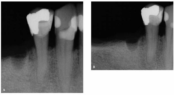
Figure 19-24A and B: (A) Pretreatment radiograph of a mandibular premolar shows one canal. (B) A second radiograph taken from an angulation of 15 degrees from the mesial discloses a second root.
Precementation Radiographs
Prior to cementation, Yamada (personal communication, 2001) re-radiographs the prepared teeth (Figures 19-25A and B). These images check the pulpal, periapical, and periodontal status of the teeth. Also, the radiographs, unencumbered by the presence of the metal castings, provide a chamber/canal road map record if the tooth requires endodontics in the future. This may appear pessimistic, but Arens and Chivian reported that over 40% of teeth requiring root canal therapy are crowned.4 Prior knowledge of the size, location, and direction of the chamber and the canal will reduce the possibility of (1) crown damage during access opening, (2) lost time searching for the canal orifice, (3) perforations of the chamber or the canal because of disorientation, (4) natural core elimination by gutting, (5) crown dislodgment, and (6) sufficient destruction to alter the situation and require corrective surgery. Each of these iatrogenic possibilities reduces the prognosis and jeopardizes the tooth's reliability as an abutment.
Diagnosis
By correlating all of the information gathered, the clinician can, within reason, determine which teeth may or may not require root canal therapy prior to the reconstructive procedures. By far the most difficult pulpal tissue status to classify is found within the confines of a previously restored tooth. For this reason, it is imperative to understand how pulps react to dental procedures.
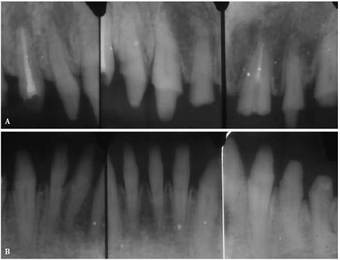
Figure 19-25A and B: Precementation radiographs provide a road map to the canals if endodontic therapy is necessary after cementation of the castings. (Radiographs courtesy of Dr. Henry Yamada.)
- on 01.11.2012 [endodontics]
- on 01.11.2012 [endodontics]
- on 01.11.2012 [endodontics]
- on 10.13.2011 [endodontics]
- on 12.15.2010 [endodontics]
- on 08.11.2010 [endodontics]
- Long Island College Hospital - [education]
- Faculty of Dental Medicine - H [education]
- The American Association of Or [organize]
- Summer Institute in Clinical D [organize]
- Academy of Osseointegration [organize]
- University of North Carolina a [education]
- American Orthodontic Society [article]
- American Equilibration Society [article]
- Niigata University - Japan [education]
- University of Buffalo [education]