ENDODONTICS AND ESTHETIC DENTISTRY(4)
Cavity Tests
When tests are inconclusive with the less apprehensive patient, drilling through the crown surface and the dentin of an unanesthetized tooth is an excellent method of investigating further if the pulp is necrotic. This should be done only if the tooth has not responded to the traditional vitality tests like cold and electric pulp testing (EPT). It must be carefully explained to the patient that based on testing, it is likely that the pulp is already necrotic; therefore, he or she should not feel any pain when the tooth is drilled. If the patient reports pain or sensitivity once the cavity preparation has reached the dentin and the "normal" response has been established with a test like the EPT, then the opening is restored. If extensive caries is present, the patient is then anesthetized, and all caries and filling materials are removed prior to restoration. This will allow visual evaluation of the cavity floor and the ability to estimate the strength of the remaining core. If the patient did not report any pain or sensation, an endodontic access is cut, and appropriate endodontic therapy is initiated after all decay has been removed from the tooth.
Electric pulp testing with a "mini-tip" through a test cavity may be the key to making a diagnosis in a tooth with a radiolucency that cannot be differentiated as either of periodontal or endodontic origin.
One can suspect a necrotic pulp if the reading is negative. Endodontics would then be the treatment of choice (Figures 19-17A, and 19-17B). If the reading is positive and there are no pulpal symptoms, periodontal therapy would be indicated (Figures 19-17C, 19-17D, and 19-17E). Endodontic treatment may be required if the root apices are compromised during periodontal procedures (Figures 19-17F, and 19-17G).
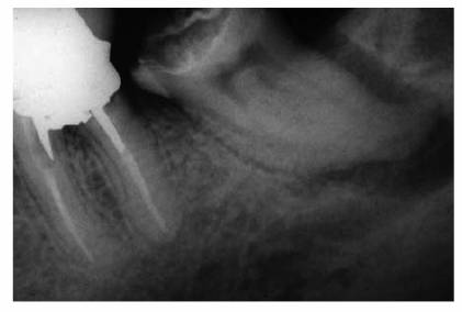
Figure 19-17A: Mandibular molar with a necrotic pulp. Root canal therapy was instituted.
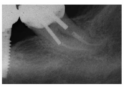
Figure 19-17B: Ten years following completion of root canal therapy there is a complete bone fill-in. No periodontal treatments were performed on this tooth.
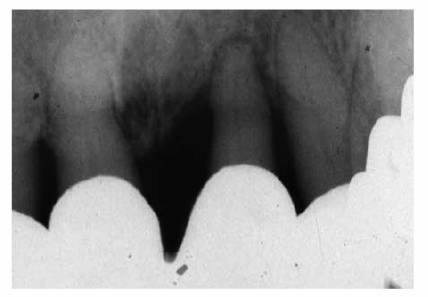
Figure 19-17C: Maxillary central incisor tooth with a vital pulp. Endodontic therapy was not indicated.
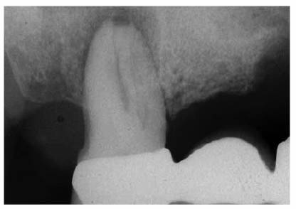
Figure 19-17D: A maxillary first molar with a periapical radiolucency.
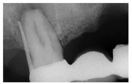
Figure 19-17E: A gutta-percha point placed in the distal pocket. Pulp testing through an occlusal opening revealed a vital pulp. The cause of the radiolucency was of periodontal origin and therapy followed that course.
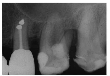
Figure 19-17F: Maxillary first molar with an uninflamed vital pulp. There was extensive bone loss surrounding the distobuccal root.
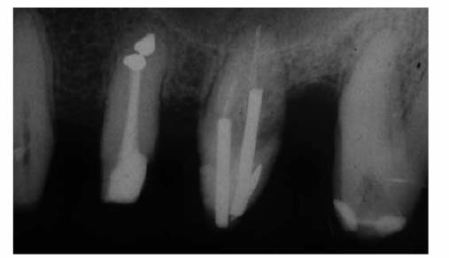
Figure 19-17G: Root canal therapy was performed to allow for the resection of the periodontally involved root.
- on 01.11.2012 [endodontics]
- on 01.11.2012 [endodontics]
- on 01.11.2012 [endodontics]
- on 10.13.2011 [endodontics]
- on 12.15.2010 [endodontics]
- on 08.11.2010 [endodontics]
- Long Island College Hospital - [education]
- Faculty of Dental Medicine - H [education]
- The American Association of Or [organize]
- Summer Institute in Clinical D [organize]
- Academy of Osseointegration [organize]
- University of North Carolina a [education]
- American Orthodontic Society [article]
- American Equilibration Society [article]
- Niigata University - Japan [education]
- University of Buffalo [education]