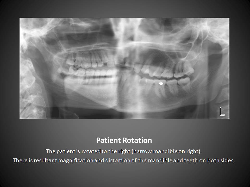
The patient must also be positioned in the centre of the focal trough. Failure of positioning may result in horizontal magnification variations, which are most marked in the anterior region of the mandible: patient positioned too far forward results in more narrow anterior teeth; patient positioned too far back results in wider anterior teeth; if the patient is rotated, the structures on the side that it is rotated to will appear smaller in width compared to the opposing side
1 / 6
Tags:
